
Assessing Keloid Risk Before Surgery: What to Know
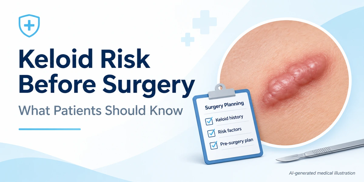
Keloid risk before surgery is evaluated by reviewing personal and family scar history, skin type, previous surgical outcomes, and the planned incision site. Clinicians use this information to estimate the likelihood of abnormal scar formation and to discuss preventive strategies. While no test can guarantee a scar‑free result, a thorough pre‑operative assessment helps tailor surgical techniques and postoperative care to minimise the chance of a keloid developing.
What Is a Keloid?
A keloid is a type of abnormal scar that grows beyond the original wound margins, often appearing raised, firm, and shiny. Unlike normal mature scars, keloids continue to produce collagen long after the wound has closed, leading to a lesion that can be larger than the initial injury. They are most common on the chest, shoulders, upper back, and earlobes, and are more prevalent in individuals with darker skin tones.
What Causes Abnormal Scarring and Keloids?
The formation of keloids involves a complex interplay of genetic, cellular, and mechanical factors. Over‑production of collagen by fibroblasts, prolonged inflammation, and increased tension on the wound edge all contribute to excessive scar tissue. Genetic predisposition, such as HLA‑DRB1*15 alleles and NEDD4 polymorphisms, has been linked to higher susceptibility pediatric scar risk model. Ethnic background, especially African, Asian, and Hispanic ancestry, also raises risk, as does a personal or family history of keloids risk assessment article.
Who Should Undergo Keloid Risk Assessment?
Anyone scheduled for a surgical procedure that creates a skin incision should be asked about prior abnormal scarring. Particular attention is given to patients who have:
- Previous keloids or hypertrophic scars.
- Close relatives with keloids.
- Fitzpatrick skin types III–VI (darker skin).
- History of acne, piercings, or trauma that healed with raised scars.
These individuals benefit from a detailed pre‑operative discussion to set realistic expectations.
How Does the Assessment Work?
Clinicians follow a stepwise approach:
1. Detailed History
Patients are asked about any past keloids, the location of those scars, and whether family members have similar lesions. The timing, type of injury, and healing course are recorded.
2. Physical Examination
The surgeon inspects the skin for existing scars, evaluates skin elasticity, and notes any areas of high tension. Certain anatomical sites—chest, shoulders, upper back, and earlobes—are recognised as higher‑risk locations risk assessment article.
3. Skin Type Evaluation
Fitzpatrick classification is used to document skin colour, as darker tones have a higher odds ratio for keloid formation (approximately 1.7 for African‑American individuals) pediatric scar risk model.
4. Use of Scoring Systems
Validated tools such as the Japanese Scar Scale (JSS) can help categorise existing scars as normal, hypertrophic, or keloid and track changes over time JSS overview. Although primarily designed for burn scars, the JSS is adapted by some clinicians for surgical scar assessment.

What Outcomes Are Realistic?
Even with optimal technique, the exact scar outcome cannot be predicted. Patients should understand that:
- Minor improvements in scar appearance are possible with tension‑reducing sutures and careful wound care.
- Complete prevention of keloid formation is not guaranteed.
- Early detection of abnormal scarring allows for timely intervention, such as silicone gel or corticosteroid injections.
Benefits and Limitations
Benefits of pre‑operative risk assessment include personalised surgical planning, informed consent, and the ability to initiate preventive measures promptly. Limitations stem from the lack of a definitive predictive test; risk models rely on population data and may not capture individual variability. Additionally, imaging modalities like ultrasound or multiphoton confocal microscopy can visualise scar tissue but cannot definitively differentiate keloids from hypertrophic scars on their own scar imaging study.
Risks or Possible Complications
The primary risk discussed during assessment is the potential development of a keloid, which can cause itching, pain, and cosmetic concern. Rarely, aggressive scar tissue can restrict movement if it forms over joints. Psychological distress related to appearance is also a recognised complication.
Follow‑Up and Monitoring
Patients identified as high risk are scheduled for early postoperative reviews, often at 2‑week intervals for the first two months. During these visits, clinicians assess scar maturation, tension, and any early signs of keloid formation. Adjustments—such as pressure therapy, silicone sheets, or early steroid injections—can be implemented based on findings.
When to Seek Medical Advice
Contact a healthcare professional promptly if you notice any of the following after surgery:
- Rapidly enlarging, raised scar that extends beyond the incision line.
- Persistent itching, tenderness, or pain in the scar area.
- Redness or thickening that does not improve after several weeks.
Early evaluation improves the chance of effective treatment.
Medical References
Jeadv Clinical Practice – Keloid Treatment Assessment Measures
Scar Formation: Biological Process and Surgical Factors – Dr Foti Sofiadellis
Keloids and Hypertrophic Scars – Dermatology Advisor
Keloid Scars: Causes, Risk Factors, Symptoms, Treatment – Continental Hospitals
Risk Assessment Model for Post‑Surgical Scar Formation – Pediatric Study
Frequently Asked Questions
How can I know if I am prone to keloids before surgery?
Your surgeon will ask about any previous keloids, family history, skin type, and the locations of past scars. These factors together help estimate your risk.
What is the difference between a keloid and a hypertrophic scar?
A keloid grows beyond the original wound margins and never regresses, while a hypertrophic scar stays within the wound boundaries and may improve over time.
Can any test definitively predict a keloid?
No single test can guarantee prediction. Risk assessment relies on clinical history, skin examination, and validated scoring tools, but individual outcomes remain uncertain.
Are there any measures that can reduce keloid formation after surgery?
Techniques such as tension-free suturing, silicone gel sheets, and early steroid injections can lower the chance of keloid development, though they cannot eliminate risk completely.
When should I contact my doctor about a postoperative scar?
If the scar becomes raised, itchy, painful, or extends beyond the incision line within a few weeks after surgery, seek medical advice promptly.
Conclusion
Assessing keloid risk before surgery involves a careful review of personal and family scar history, skin type, and the planned incision site. While clinicians can identify high‑risk factors and recommend preventive strategies, no method guarantees a scar‑free outcome. Understanding the limits of prediction helps patients set realistic expectations and seek timely care if abnormal scarring develops.
Medical Disclaimer
This article is intended for general educational purposes only and does not replace individual medical advice, diagnosis or treatment. Suitability, risks and outcomes vary. Consult a qualified healthcare professional for medical guidance specific to your situation.