
Trichoscopy for Hair Loss: How Scalp Examination Improves Diagnosis
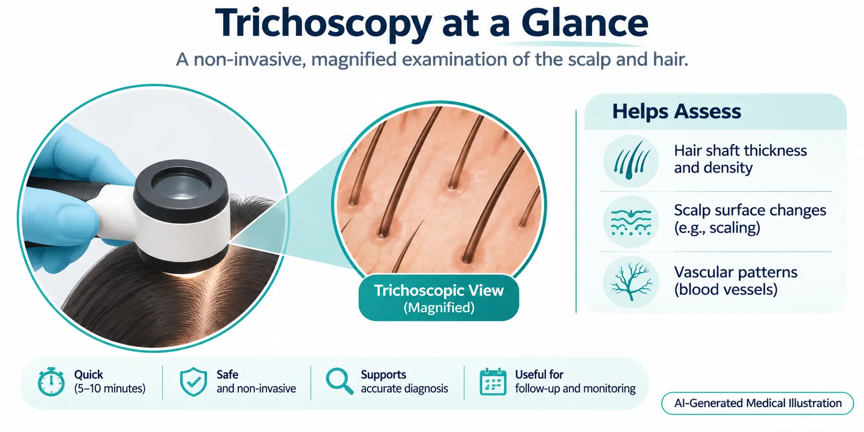
Trichoscopy is a non‑invasive, magnified examination of the scalp and hair shafts performed with a handheld dermatoscope. By visualising individual follicles, scaling, and vascular patterns, clinicians can differentiate common causes of hair loss without needing a biopsy. The technique provides immediate visual feedback, supports a more accurate differential diagnosis, and helps decide whether a patient is a candidate for hair‑restorative procedures such as transplantation. It is safe, quick, and can be repeated to monitor treatment response.
What Is Trichoscopy?
Trichoscopy, also called scalp dermoscopy, uses polarized or non‑polarized light to enlarge the view of hair and scalp structures up to 70×. The method reveals details invisible to the naked eye, such as perifollicular erythema, black dots, exclamation‑mark hairs, and the presence of vellus versus terminal hairs. These visual clues are essential for classifying alopecia types.
Why Is Trichoscopy Performed?
The primary goal is to obtain a rapid, visual diagnosis of hair‑loss disorders while avoiding invasive procedures. It assists clinicians in distinguishing inflammatory alopecia (e.g., alopecia areata, lichen planopilaris) from non‑inflammatory patterns (e.g., androgenetic alopecia, telogen effluvium). Early identification can guide timely treatment and improve outcomes.
Who May Need Trichoscopy?
Anyone presenting with noticeable thinning, patchy loss, or diffuse shedding may benefit. It is especially useful for:
- Patients with unclear etiology after history and basic examination.
- Individuals considering hair‑transplant surgery, where donor‑site quality must be assessed.
- Clients in salon settings who seek professional advice on scalp health.
For a broader view of diagnostic steps, see our Hair Pull Test for Hair Loss.
How Does Trichoscopy Work?
The clinician places the dermatoscope directly on the scalp, often after a brief cleaning to remove sebum and product residue. Light and magnification reveal:
- Follicular openings and their density.
- Hair shaft thickness and shape.
- Scalp surface features such as scaling, crusting, or vascular changes.
Images can be captured for documentation and compared over time. The process typically takes 5–10 minutes and does not require anesthesia.

What Do Trichoscopic Findings Mean?
Specific patterns correlate with particular conditions:
- Yellow dots – often seen in androgenetic alopecia and alopecia areata.
- Black dots – indicate broken hairs at the scalp surface, common in alopecia areata.
- Perifollicular scaling – suggests inflammatory disorders such as seborrheic dermatitis or psoriasis.
- Vellus‑hair predominance – points toward miniaturisation in androgenetic alopecia.
Interpretation requires clinical context; findings alone cannot provide a definitive diagnosis.
Benefits and Limitations
Benefits include immediate visual feedback, non‑invasiveness, low cost, and suitability for repeated monitoring. Limitations involve operator dependence, the need for training to recognise subtle patterns, and the inability to detect deep dermal pathology that may require a biopsy.
Risks or Possible Complications
Trichoscopy is considered safe. Minor discomfort may occur if the device contacts a highly sensitive scalp, and very rare allergic reactions to contact gels are possible. There are no known long‑term adverse effects.
Recovery or Follow‑Up
There is no recovery period; patients can resume normal activities immediately. Follow‑up examinations are typically scheduled every 3–6 months to track disease progression or treatment response. Documentation of serial images enhances objective assessment.
When to Seek Medical Advice
If you notice rapid hair loss, scalp pain, itching, or visible lesions, schedule a professional evaluation. Trichoscopy can be part of that assessment, but persistent or worsening symptoms may require additional tests such as blood work or scalp biopsy.
Medical References
Scalp Smart Pro – Hair + Scalp Education for Stylists
Hair & Scalp Diagnostic Consultations – A Mé Vitality overview
Hair Care Professionals as Community Health Partners in Detection and Management of Alopecia
Frequently Asked Questions
1. What equipment is needed for trichoscopy?
A handheld dermatoscope with polarized or non-polarized light, optional contact gel, and a camera for image capture are the core components.
2. Can trichoscopy replace a scalp biopsy?
No. Trichoscopy can suggest a diagnosis, but a biopsy may still be required for definitive histopathological confirmation in ambiguous cases.
3. Is trichoscopy painful?
The procedure is generally painless; mild pressure may be felt if the scalp is particularly sensitive, but no anesthesia is needed.
4. How often should trichoscopy be performed?
Routine monitoring every 3–6 months is common, though frequency depends on the underlying condition and treatment plan.
5. Can trichoscopy be done in a salon?
Yes, many trained stylists use dermatoscopes for basic scalp assessment, but any concerning findings should be referred to a dermatologist.
6. What are common trichoscopic signs of androgenetic alopecia?
Typical signs include hair-shaft miniaturisation, increased vellus-hair proportion, and yellow dots within follicular openings.
7. Does trichoscopy help determine hair-transplant candidacy?
It assists by evaluating donor-site density and scalp health, which are key factors in planning a successful transplant.
Conclusion
Trichoscopy offers a quick, non‑invasive window into scalp and hair‑shaft health, helping clinicians differentiate among common alopecia types and assess suitability for procedures like hair transplantation. While it provides valuable visual clues, interpretation must be paired with a thorough history and, when necessary, further testing. Understanding its benefits and limits empowers patients and professionals to make informed decisions about hair‑loss management.
Medical Disclaimer :
This article is intended for general educational purposes only and does not replace individual medical advice, diagnosis or treatment. Suitability, risks and outcomes vary. Consult a qualified healthcare professional for medical guidance specific to your situation.