
Nasal Septum Anatomy: Structure, Function & Breathing Role
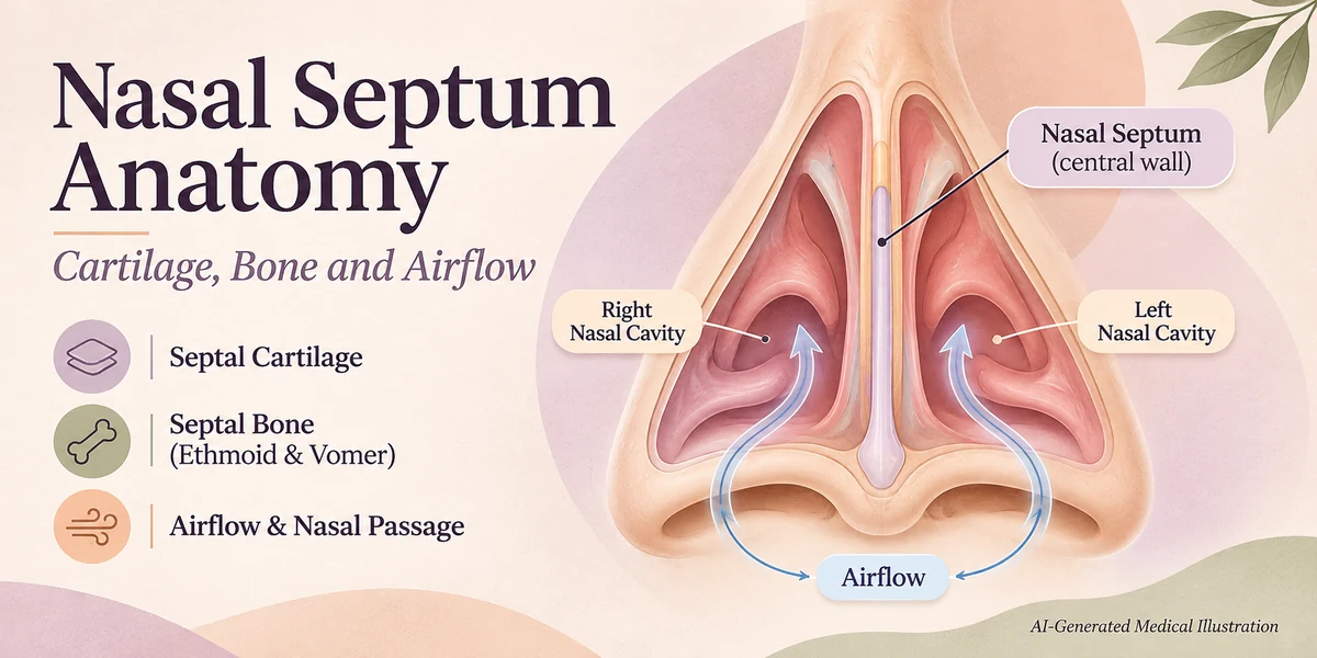
The nasal septum is the central wall that separates the right and left nasal cavities, providing structural support for the nose and helping to regulate airflow. It is composed of both cartilage and bone, covered by a moist mucosal lining that warms and filters inhaled air. By forming the medial wall of each nasal passage, the septum works with the nasal valves, turbinates and mucosa to maintain smooth, symmetrical breathing. Understanding its anatomy clarifies why some people experience nasal obstruction and when specialist assessment may be needed.
What Is the Nasal Septum?
The nasal septum is a thin, yet sturdy, partition that divides the nasal cavity into two symmetrical chambers. It creates the medial boundary of each cavity and contributes to the external shape of the nose. The septum is covered on both sides by a vascular mucous membrane that humidifies, warms, and filters the air we breathe anatomy.co.uk.
What Is Its Structure?
The septum consists of three main components:
- Quadrangular (septal) cartilage: A flexible hyaline cartilage sheet that forms the anterior two‑thirds of the septum and extends from the columella to the junction with the bony parts.
- Perpendicular plate of the ethmoid bone: The superior‑posterior bony plate that articulates with the cartilage and forms the upper portion of the septum.
- Vomer: The inferior‑posterior bone that meets the perpendicular plate at an oblique angle, completing the posterior septal wall.
These elements interlock to create a rigid yet slightly flexible framework that supports the nose and separates the airflow streams. Detailed descriptions of each component are provided by Kenhub and IntechOpen.
Why Is the Septum Important for Breathing?
The septum works together with the internal nasal valves, turbinates, and mucosal lining to direct airflow. A straight, well‑positioned septum ensures that air passes evenly through both cavities, allowing optimal humidification and resistance. When the septum deviates, it can narrow one side of the airway, increasing turbulent flow and potentially causing unilateral obstruction, a sensation of “blocked” breathing, or difficulty sleeping.
Who May Experience Septal Deviation?
Most people have some degree of anatomical variation in septal alignment. A clinically significant deviation—one that causes symptoms such as persistent nasal obstruction, recurrent sinusitis, or epistaxis—occurs in a minority of individuals. Not every deviation warrants treatment, and many remain asymptomatic throughout life.
How Does the Septum Influence Airflow?
Air enters the nostrils and is guided by the septum toward the nasal valve region, the narrowest part of the airway. The turbinates, attached to the lateral walls, further condition the air. If the septum is deviated, the cross‑sectional area on the affected side is reduced, increasing resistance according to Poiseuille’s law. This can lead to a sensation of difficulty breathing through the nose, especially when lying down.
What Do Clinical Findings Mean?
Evaluation begins with an anterior rhinoscopy—visual inspection using a speculum and light—to assess septal alignment, mucosal health, and the presence of crusting or bleeding. If deeper structures need assessment, nasal endoscopy provides a detailed view of the posterior septum, turbinates, and sinus openings. Imaging (CT or MRI) is reserved for complex cases, such as suspected sinus disease, trauma, or when surgical planning is required PMC review.
Benefits and Limitations of Surgical Intervention
When a deviation causes significant symptoms, septoplasty can reposition or remove excess cartilage and bone to improve airway patency. Septorhinoplasty combines septal correction with cosmetic reshaping of the nasal tip or dorsum, while rhinoplasty alone addresses appearance without correcting the septum. Surgical outcomes vary; many patients report improved breathing, but results are not guaranteed, and some may retain residual obstruction.
Risks or Possible Complications
All nasal surgeries carry potential risks, including bleeding, infection, septal perforation, altered sensation, or unsatisfactory cosmetic change. Specific to septal work, a perforation can create whistling sounds during breathing. Patients should discuss these possibilities and obtain informed consent Understanding Surgical Consent Forms: Risks & Rights.
When to Seek Medical Advice
Consider professional evaluation if you experience persistent unilateral nasal blockage, frequent nosebleeds, chronic sinus infections, or sleep‑related breathing difficulties that do not improve with medical therapy (e.g., nasal steroids or saline irrigation). An ENT specialist can determine whether anatomical factors, such as a deviated septum, contribute to symptoms and recommend appropriate management.

Comparison of Common Nasal Procedures
| Procedure | Primary Goal | Typical Indications | Key Risks |
|---|---|---|---|
| Septoplasty | Correct septal deviation to improve airflow | Symptomatic deviation, recurrent sinusitis | Bleeding, perforation, altered sensation |
| Rhinoplasty | Alter external nasal shape for cosmetic reasons | Patient‑desired aesthetic change | Unsatisfactory appearance, breathing changes |
| Septorhinoplasty | Combine functional septal correction with cosmetic reshaping | Deviated septum plus aesthetic concerns | Combined risks of both procedures |
Medical References
Frequently Asked Questions
What is the nasal septum made of?
The septum consists of quadrangular cartilage at the front, the perpendicular plate of the ethmoid bone superiorly, and the vomer bone inferiorly, all covered by a moist mucosal lining.
Does every deviated septum cause breathing problems?
No. Many people have slight deviations that never produce symptoms. Only clinically significant deviations that narrow the airway may lead to obstruction or related issues.
How can I tell if my nasal obstruction is due to a deviated septum?
Persistent unilateral blockage, frequent nosebleeds, or difficulty breathing that does not improve with medical treatment may suggest a septal deviation, but an ENT examination is needed for confirmation.
What is the difference between septoplasty and rhinoplasty?
Septoplasty corrects the internal cartilage and bone to improve airflow, while rhinoplasty reshapes the external nose for cosmetic reasons. Septorhinoplasty combines both goals.
What are the main risks of septal surgery?
Potential complications include bleeding, infection, septal perforation, altered sensation, and unsatisfactory cosmetic outcomes.
Can a deviated septum cause sinus infections?
A significant deviation can impede sinus drainage, increasing the risk of recurrent sinusitis, but other factors also contribute to infection.
When should I see an ENT specialist about my nose?
If you have ongoing nasal blockage, frequent nosebleeds, chronic sinus issues, or sleep-related breathing problems that persist despite medical therapy, an ENT referral is advisable.
Conclusion
The nasal septum is a crucial anatomical structure that not only supports the external nose but also ensures balanced airflow through the nasal passages. While minor deviations are common and often harmless, significant misalignments can affect breathing and may require professional assessment. Understanding the septum’s components, function, and the realistic expectations of surgical options empowers individuals to make informed decisions about their nasal health.
Medical Disclaimer
This article is intended for general educational purposes only and does not replace individual medical advice, diagnosis or treatment. Suitability, risks and outcomes vary. Consult a qualified healthcare professional for medical guidance specific to your situation.