
Otoplasty in Navi Mumbai: Scars, Recovery & Natural Results
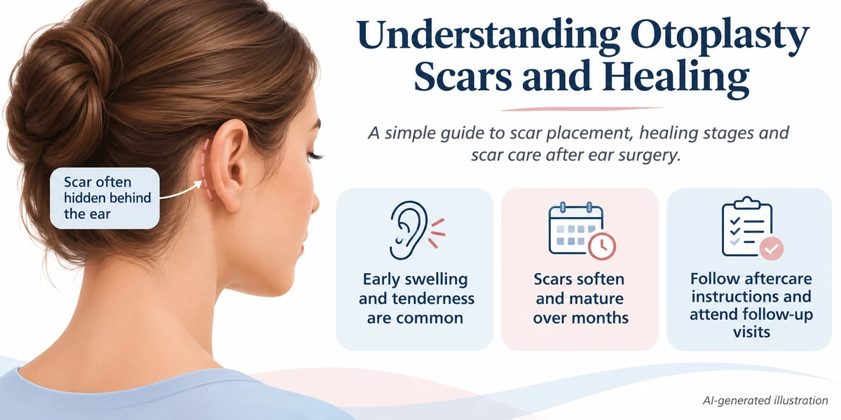
Otoplasty Scars and Recovery: Healing, Aftercare and Natural Results
Otoplasty scars are usually located behind the ears or within natural ear folds, but they are permanent and do not disappear completely. Recovery after otoplasty, also called ear pinning or ear reshaping surgery, commonly involves temporary swelling, bruising, tenderness and protective dressings. Many people resume routine activities within one to two weeks, while deeper healing and scar maturation can take several months.
Otoplasty changes the shape, position or proportion of the external ear. It may be considered for prominent ears, ear asymmetry or certain structural differences.
Although scars may be relatively inconspicuous, their appearance depends on the surgical technique, skin characteristics and individual wound healing.
This article explains what otoplasty scars may look like, how recovery progresses, when the ears may appear more natural and which symptoms require medical assessment.
What Is Otoplasty?
Otoplasty is a surgical procedure that changes the appearance or structure of the outer ear.
It may involve:
Reshaping ear cartilage
Creating or modifying natural ear folds
Repositioning prominent ears
Correcting selected asymmetries
Adjusting ear size or proportions in appropriate cases
Addressing certain congenital or injury-related deformities
Ear pinning typically focuses on repositioning prominent ears closer to the head.
Other forms of otoplasty may involve more extensive cartilage or tissue reconstruction.
Cosmetic otoplasty does not generally improve hearing, because it primarily changes the external ear rather than the structures responsible for hearing.
Where Are Otoplasty Scars Located?
The location of an otoplasty scar depends on the technique used.
1. Behind-the-Ear Incision
A common approach uses an incision behind the external ear, where the ear meets the side of the head.
This location may make the scar less noticeable during ordinary viewing.
However, the scar is still present and may be visible when the ear is examined closely.
2. Incisions Within Ear Folds
Some procedures use incisions in or near natural folds of the external ear.
The final scar location depends on the cartilage changes required.
3. Additional Incisions for Complex Surgery
Certain reconstructive or ear reduction procedures may require different incision patterns.
More extensive operations can involve additional scars.
The surgeon should explain the proposed incision locations before treatment.
Are Otoplasty Scars Permanent?
Yes. Every surgical incision produces a scar.
Scars commonly change in appearance as the wound heals and collagen remodels.
Early scars may appear:
Pink or red
Slightly raised
Firm
Sensitive
More noticeable than expected
Over time, some scars become flatter, softer and less conspicuous.
However, scar maturation is individual.
A scar may remain visible, become thickened or develop an abnormal appearance.
No otoplasty technique guarantees invisible or scar-free healing.
How Do Otoplasty Scars Change Over Time?
Scar appearance evolves gradually.
| Healing stage | Possible scar changes |
|---|---|
| First 1–2 weeks | Incision healing, tenderness and possible redness |
| Weeks 3–6 | Early scar formation and gradual reduction in wound sensitivity |
| Months 2–3 | Scar may begin softening as tissue healing continues |
| Months 3–6 | Further changes in colour, firmness and visibility |
| Months 6–12 | Continued scar maturation |
| Beyond 12 months | Some scars may continue changing, while others remain noticeable |
These are approximate stages rather than guaranteed outcomes.
Some people develop hypertrophic scars or keloids, particularly when they are predisposed to abnormal scar formation.
What Factors Affect Otoplasty Scar Appearance?
Several factors influence how a scar heals.
Surgical Technique
Incision placement, tissue handling and wound closure can affect the eventual appearance.
Individual Skin Characteristics
Skin type and a personal or family history of abnormal scarring may be relevant.
Wound Healing
Infection, wound separation or excessive tension can interfere with scar formation.
Pressure or Injury
Repeated friction or accidental pulling during early recovery may disturb the incision.
Smoking and Nicotine Exposure
Smoking and nicotine can impair wound healing and increase certain postoperative risks.
Previous Surgery
Earlier incisions or scar tissue may influence subsequent healing.
These factors do not allow a clinician to predict exactly how any individual scar will look.
How Is Otoplasty Surgery Performed?
The precise technique depends on ear anatomy and the intended correction.
Step 1: Medical and Ear Assessment
The surgeon reviews medical history, medication, allergies, ear structure and the reasons for considering surgery.
Step 2: Anaesthesia
Local anaesthesia, sedation or general anaesthesia may be selected according to age, medical needs and procedure complexity.
Step 3: Incision and Cartilage Access
An incision is commonly made behind the ear or within an ear fold.
Step 4: Cartilage Reshaping
The cartilage may be folded, repositioned, modified or secured using sutures.
Step 5: Wound Closure
The incision is closed with sutures.
Some sutures are absorbable; others may require removal.
Step 6: Protective Dressing
A dressing or bandage may be applied to protect the ears and support the early postoperative position.
For a more detailed explanation of surgical methods, read the Otoplasty Procedure and Ear Correction Guide.
How Long Does Otoplasty Recovery Take?
Initial recovery often takes approximately one to two weeks, but healing is not complete when ordinary activities resume.
Swelling, stiffness and tenderness may continue improving over subsequent weeks.
The timing depends on:
The surgical technique
Extent of cartilage modification
Patient age and medical health
Wound healing
Dressing requirements
Postoperative complications
Individual recovery response
More extensive reconstruction can require a different recovery schedule.
Otoplasty Recovery Timeline
| Recovery period | What may happen | General care considerations |
|---|---|---|
| First 24–72 hours | Swelling, soreness, bruising or tightness | Rest, protect the ears and follow medication instructions |
| Days 4–7 | Early discomfort may improve; dressings may be reviewed | Keep dressings intact as instructed |
| Weeks 1–2 | Gradual return to some routine activities | Avoid pressure, pulling and accidental ear injury |
| Weeks 2–4 | Swelling and bruising often continue settling | Follow headband and activity instructions |
| Weeks 4–6 | Further healing and reduced tenderness | Resume physical activities only when cleared |
| Weeks 6–8 | Many individuals experience more complete initial healing | Continue protecting the ears when advised |
| Months 3–6 | Ear contour and scar appearance may continue changing | Monitor scar maturation and attend follow-up |
| Months 6–12 | Further scar softening and tissue changes may occur | Seek assessment for persistent concerns |
These timeframes are general estimates, not fixed milestones.
What Is Normal During the First Week After Otoplasty?
Several temporary symptoms can occur.
Swelling
Swelling develops as part of the normal tissue-healing response.
The ears may initially appear larger or more prominent than expected.
Bruising
Bruising may develop around the ears or nearby skin.
Tenderness
The ears may feel sore or sensitive when touched.
Tightness
The cartilage, skin or surrounding tissue may feel tight because of swelling and surgical repositioning.
Temporary Numbness
Altered skin sensation can occur during early healing.
Itching Beneath Dressings
Some people experience itching under protective bandages.
Do not remove dressings or scratch healing incisions without appropriate medical instructions.
Symptoms should generally move towards improvement rather than becoming progressively more severe.
Why Are Bandages Used After Otoplasty?
Bandages or dressings may be used to:
Protect the surgical incisions
Reduce accidental trauma
Support the ears during early healing
Limit unnecessary movement
Help maintain the planned postoperative position
The type and duration of dressing use depend on the procedure.
Removing dressings prematurely may disturb healing.
Follow the surgeon’s specific instructions about changing or removing bandages.
Do You Need to Wear a Headband After Otoplasty?
Some patients are advised to wear a protective headband, particularly during sleep.
The purpose may be to reduce accidental folding, pressure or injury to the ears.
However, the duration of use varies.
A headband should not be used as a substitute for the prescribed surgical dressing.
Do not wear excessively tight headwear or change the recommended schedule without advice from the treating team.
How Should You Sleep After Otoplasty?
Sleeping positions may need temporary modification to avoid pressure on healing ears.
General recommendations may include:
Sleeping on the back during early recovery
Avoiding direct pressure on the operated ear
Using a prescribed protective headband
Avoiding sleeping positions that fold or pull the ear
Keeping dressings in place as instructed
The exact recommendations depend on the technique and healing progress.
When Can You Return to Work or School?
Many individuals can return to light work or school activities within approximately one to two weeks, depending on comfort and medical clearance.
However, the appropriate timing depends on the activity.
| Activity | Recovery consideration |
|---|---|
| Desk-based work | May be resumed when comfortable and medically appropriate |
| School attendance | Depends on healing and ability to avoid ear injury |
| Light walking | Often possible relatively early |
| Heavy physical work | May require a longer restriction period |
| Contact sports | Usually restricted until adequate healing and medical clearance |
| Activities involving helmets | Pressure on the ears requires particular consideration |
| Swimming | Should be delayed until wound healing and medical clearance |
No universal return-to-activity date applies to every patient.
When Can You Exercise After Otoplasty?
Physical activity should resume gradually.
Strenuous exercise may increase swelling, while contact sports can cause direct injury to healing cartilage.
During early recovery, avoid activities that:
Cause pressure on the ears
Require tight protective equipment
Involve significant risk of impact
Pull or bend the external ear
The treating surgeon should determine when specific activities can safely resume.
When Will Otoplasty Results Look Natural?
Changes in ear position may become visible after the initial dressings are removed.
However, early swelling and stiffness can make the appearance temporarily different from the eventual contour.
As healing progresses:
Swelling may gradually decrease
Ear contours may become clearer
The tissues may soften
Incision scars may mature
The position of the ears may settle
Natural-looking results are not guaranteed.
A person’s perception of natural ear shape depends on individual anatomy, cartilage characteristics, surgical technique and healing.
What Does a Natural-Looking Otoplasty Result Mean?
A natural-looking appearance generally refers to an ear contour that does not appear unnaturally flattened, sharply folded or excessively positioned against the head.
The surgeon may consider:
The relationship between the ear and scalp
Ear cartilage folds
The upper, middle and lower ear contours
Differences between the two ears
Overall facial proportions
Natural variation in ear anatomy
However, the ears are not usually identical in shape or position before surgery.
Perfect symmetry is not a realistic requirement or a guaranteed surgical outcome.
Can Otoplasty Be Overcorrected?
Yes. Excessive repositioning or cartilage modification may create an unnatural contour.
Possible concerns include:
Ears appearing too close to the head
Unusual cartilage folds
Excessively sharp contours
Uneven ear positioning
Differences between the upper and lower parts of the ear
Surgical revision may sometimes be considered, but further surgery also involves risks.
Can Ears Become Prominent Again After Otoplasty?
Yes. Some degree of recurrence is possible.
Possible contributing factors include:
Cartilage characteristics
Suture-related problems
Healing changes
Accidental trauma
Differences in surgical technique
Recurrence does not occur in every patient.
However, otoplasty cannot guarantee that the ear will remain in exactly the same position throughout life.
What Are the Possible Risks and Complications of Otoplasty?
Otoplasty is a surgical procedure and carries recognised complications.
Bleeding and Hematoma
Blood may collect beneath the skin, causing swelling, pressure and pain.
Infection
The surgical incision or surrounding tissue can become infected.
Infection involving ear cartilage requires prompt medical assessment.
Poor Wound Healing
The incision may heal slowly or partially separate.
Abnormal Scarring
Hypertrophic scars or keloids can occur in predisposed individuals.
Changes in Skin Sensation
Numbness, increased sensitivity or other sensory changes may develop.
Some changes may persist.
Persistent Asymmetry
Differences in ear position or contour may remain following surgery.
Overcorrection or Undercorrection
The resulting ear position may differ from the planned correction.
Suture Problems
Some patients may experience discomfort, suture exposure or other suture-related complications.
Recurrence
The ear may partially return towards its original position.
Need for Revision Surgery
A further operation may occasionally be considered.
However, revision surgery carries additional risks and cannot guarantee complete correction.
Normal Recovery vs Warning Signs
| May occur during recovery | Requires medical assessment |
|---|---|
| Mild swelling that gradually improves | Rapidly increasing swelling |
| Temporary soreness | Severe or worsening pain |
| Bruising that slowly settles | Expanding bruising with pressure or significant pain |
| Temporary numbness | New or worsening concerning sensory changes |
| Mild itching beneath a dressing | Increasing redness, warmth or discharge |
| Incision tenderness | Wound separation or persistent bleeding |
| Gradual scar formation | Increasingly raised, painful or abnormal scarring |
Seek urgent medical attention for severe pain, rapidly increasing swelling, persistent bleeding, high fever or signs of significant infection.
Breathing difficulty or symptoms of a severe allergic reaction require emergency care.
How Can You Care for Otoplasty Scars?
Scar care should begin only when medically appropriate.
Keep the Incision Clean
Follow the surgeon’s cleaning instructions and avoid unnecessary handling.
Protect the Wound
Avoid accidental pulling, scratching or pressure.
Do Not Remove Sutures Yourself
Suture removal, when necessary, should be performed according to medical instructions.
Discuss Silicone Scar Products
Silicone gel or silicone sheets may be considered for selected healed scars.
They should not be applied to an open wound unless specifically advised.
Avoid Unapproved Scar Massage
Scar massage may not be appropriate during early healing.
Begin only if the treating clinician recommends it and confirms that the wound has healed sufficiently.
Protect Healing Skin
Sun protection may help reduce unwanted pigmentation changes when the area is exposed.
Monitor Abnormal Scarring
A scar that becomes increasingly raised, thick, painful or itchy may require assessment.
No cream, sheet or massage technique can guarantee an invisible scar.
Otoplasty for Adults vs Children: Does Recovery Differ?
Otoplasty can be performed in appropriately selected adults and children.
The healing process may involve similar concerns, including swelling, bruising, bandages and activity restrictions.
However, children may need additional assistance with:
Avoiding accidental ear injury
Keeping dressings in place
Wearing protective headwear when advised
Following school and sports restrictions
Communicating pain or discomfort
Age alone does not determine how quickly an individual will heal.
For a detailed comparison, read Otoplasty for Adults vs Children: What Is Different?.
Otoplasty Scars and Recovery: Safety Checklist

Before surgery and during recovery, review the following:
Discuss incision placement: Understand where permanent scars may develop.
Review medical suitability: Disclose health conditions, allergies and previous ear procedures.
Understand possible complications: Include infection, hematoma, scarring, asymmetry and recurrence.
Keep dressings intact: Follow instructions about changing or removing bandages.
Protect the ears while sleeping: Avoid pressure or folding during early healing.
Use a headband only as advised: Follow the prescribed schedule and fit.
Maintain appropriate wound care: Clean incisions only as instructed.
Avoid trauma: Limit activities that could bend or injure healing cartilage.
Delay strenuous activity: Resume exercise when medically cleared.
Do not begin scar treatment prematurely: Discuss silicone products and massage before use.
Attend follow-up visits: Allow healing and cartilage position to be assessed.
Recognise warning signs: Report severe pain, rapidly increasing swelling, infection or persistent bleeding.
These precautions support recovery but cannot eliminate the possibility of complications.
Frequently Asked Questions
1. Are otoplasty scars visible?
Otoplasty scars are often placed behind the ear or within natural folds, making them less noticeable. However, scars are permanent and may remain visible or become thickened.
2. How long does it take for otoplasty scars to fade?
Scars may gradually soften and become less noticeable over several months. Scar maturation can continue for a year or longer, and complete disappearance cannot be guaranteed.
3. How long does recovery take after otoplasty?
Many individuals resume routine activities within one to two weeks. More complete healing often takes several additional weeks, while scar maturation may continue for months.
4. Do I need to wear a headband after otoplasty?
Some patients are advised to wear a protective headband, particularly while sleeping. The duration depends on the procedure and the treating surgeon's instructions.
5. When can I sleep on my side after otoplasty?
Side sleeping may be restricted during early recovery to prevent pressure or injury to the operated ear. Resume it according to medical clearance.
6. Will my ears look natural after otoplasty?
Otoplasty may improve selected concerns involving ear position or shape, but natural-looking results and perfect symmetry cannot be guaranteed. The final appearance depends on anatomy, technique and healing.
7. What are the warning signs after otoplasty?
Severe or worsening pain, rapidly increasing swelling, persistent bleeding, fever, spreading redness, discharge or wound separation require medical assessment.
Conclusion
Otoplasty scars and recovery vary according to the surgical technique, ear cartilage characteristics and individual healing.
Incisions are commonly positioned behind the ears or within natural folds, but permanent scars remain.
Swelling, bruising, tenderness and temporary numbness may occur during early recovery. Routine activities often resume within the first few weeks, while scar maturation and tissue changes may continue for several months.
Appropriate dressing care, protection from pressure, activity restrictions and postoperative follow-up can support healing.
However, no procedure guarantees invisible scars, perfectly symmetrical ears or a particular cosmetic result.
Disclaimer: All content and visuals on this website are for educational and informational purposes only. Images may be AI-generated or illustrative. Individual treatment results may vary. This information does not replace professional medical advice, diagnosis or consultation.