
Nasal Valve Function: Pre‑Rhinoplasty Breathing Assessment
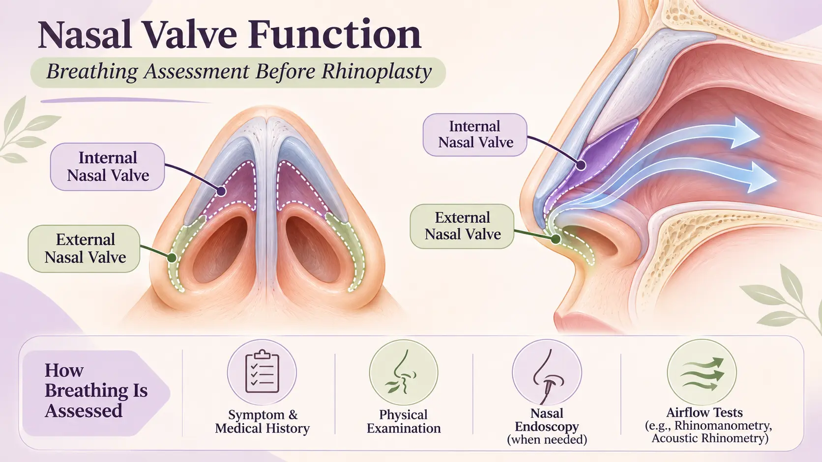
Nasal valve function refers to how well the narrow, cartilage-supported regions near the entrance of the nose allow air to pass during breathing. Before rhinoplasty, clinicians may assess nasal valve structure and movement, review breathing symptoms, examine the septum and turbinates, and use additional tests when appropriate.
A nasal valve can be narrowed at rest or collapse inward during inhalation. However, nasal obstruction has several possible causes, and no single symptom or examination manoeuvre establishes every diagnosis. Understanding how the nasal valve is assessed helps patients participate in informed discussions about treatment options and their limitations.
What Is the Nasal Valve and Why Is It Important for Breathing?
The nasal valve describes areas near the front of the nasal airway that contribute substantially to airflow resistance.
It is not a separate organ. Instead, it is formed by neighbouring cartilage, soft tissue and nasal structures working together.
The nasal valve is commonly divided into two anatomical regions:
Internal nasal valve: Located inside the nose, near the junction between the upper lateral cartilage and nasal septum. It is an important narrow segment of the nasal airway.
External nasal valve: Located closer to the nostril opening and supported by the lower lateral cartilage, surrounding soft tissue and adjacent structures.
Both regions must maintain sufficient space and structural support during breathing.
When the nasal sidewall is weak or the passage is excessively narrow, airflow may become restricted.
For more information about neighbouring structures, read Nasal Septum Anatomy: Structure, Function and Breathing.
What Is the Difference Between Internal and External Nasal Valve Dysfunction?
The internal and external nasal valves occupy different parts of the nasal airway. Problems affecting either region may cause obstruction, but the underlying anatomical mechanism can differ.
| Feature | Internal Nasal Valve | External Nasal Valve |
|---|---|---|
| Location | Narrow region inside the anterior nasal passage | Region around the nostril entrance |
| Main structures | Upper lateral cartilage, septum and nearby turbinate region | Lower lateral cartilage, nostril rim and surrounding tissues |
| Possible dysfunction | Narrowing or weakness of the internal sidewall | Nostril narrowing or inward movement of the rim |
| Clinical examination | Internal inspection and assessment during breathing | External observation of the nostril during inhalation |
| Main limitation | Symptoms may overlap with septal deviation | Collapse may be absent during quiet breathing |
These differences help clinicians identify which structures may be contributing to nasal obstruction.
However, internal and external valve problems can coexist, and neither can be diagnosed reliably through appearance alone.
What Is Static vs Dynamic Nasal Valve Collapse?
Nasal valve dysfunction can be classified as static or dynamic.
Static Nasal Valve Narrowing
Static narrowing means the airway is structurally restricted even when the person is not taking a deep breath.
Possible contributing factors include:
Naturally narrow nasal anatomy
Previous injury
Postoperative scar tissue
Changes in cartilage position
Associated septal deviation
Dynamic Nasal Valve Collapse
Dynamic collapse occurs when part of the nasal sidewall moves inward during inhalation.
During breathing in, pressure changes within the nasal passage can pull an inadequately supported sidewall toward the centre of the airway.
Symptoms may become more noticeable during deep breathing or physical activity.
Importantly, a person may have both static narrowing and dynamic collapse.
Distinguishing these conditions can influence clinical assessment and treatment planning.
What Symptoms May Suggest Nasal Valve Dysfunction?
Nasal valve dysfunction may be associated with:
Persistent difficulty breathing through one or both nostrils
Nasal blockage that becomes more noticeable during deep inhalation
Visible inward movement of the nostril or nasal sidewall
Difficulty breathing through the nose during exercise
A sensation of inadequate airflow despite previous nasal treatment
Temporary improvement when the nasal sidewall is gently supported
These symptoms are not exclusive to nasal valve dysfunction.
Allergic rhinitis, enlarged turbinates, septal deviation, nasal polyps and other conditions can produce similar complaints.
Therefore, symptoms should be considered alongside medical history and clinical examination.
How Is Nasal Breathing Evaluated Before Rhinoplasty?
A preoperative nasal breathing assessment helps clinicians identify possible causes of obstruction and determine whether functional concerns should influence surgical planning.
1. Medical History and Symptom Assessment
The clinician may ask:
Is breathing difficulty present on one side or both?
Does the obstruction change throughout the day?
Is breathing worse during exercise, sleep or deep inhalation?
Is there a history of nasal injury?
Has the patient undergone previous septoplasty or rhinoplasty?
Are allergies, sinus symptoms or frequent congestion present?
Have previous medical treatments helped?
These questions help distinguish persistent structural obstruction from symptoms that fluctuate with inflammation or other conditions.
2. External Nasal Examination
The clinician examines the outside of the nose and observes the nostrils during quiet and deeper breathing.
This may identify:
Narrow nostril openings
Inward movement of the nasal sidewall
Asymmetry
Reduced cartilage support
Changes following previous nasal surgery
A normal appearance at rest does not completely exclude dynamic nasal valve dysfunction.
3. Internal Nasal Examination
Anterior rhinoscopy allows clinicians to inspect the front of the nasal passages.
The examination can identify septal abnormalities, turbinate enlargement, mucosal inflammation and visible anatomical narrowing.
4. Additional Examination When Needed
Nasal endoscopy or supplementary investigations may be considered when the diagnosis remains uncertain or other nasal conditions are suspected.
The assessment should be individualised rather than based on a routine requirement for every available test.
What Is the Cottle Maneuver?
The Cottle maneuver is a clinical examination technique in which a clinician gently pulls the cheek outward to assess whether perceived nasal breathing improves.
The manoeuvre temporarily widens portions of the anterior nasal airway.
Improved airflow sensation may suggest that widening the nasal sidewall provides relief.
However, the test is not sufficiently specific to identify the exact site or cause of obstruction by itself.
A positive result should therefore be interpreted with the patient’s symptoms and other clinical findings.
What Is the Modified Cottle Maneuver?
The modified Cottle maneuver involves selectively supporting specific regions of the nasal sidewall while assessing a person’s breathing sensation.
Unlike the standard Cottle maneuver, it can help clinicians investigate different areas of possible narrowing.
For example, the examiner may support the internal or external nasal valve region separately.
This can provide useful information about where additional structural support might influence airflow.
Nevertheless, improvement during the test does not guarantee that surgical treatment will be beneficial.
Clinical findings must be interpreted together rather than allowing one manoeuvre to determine the treatment plan.
What Is the NOSE Score?
The Nasal Obstruction Symptom Evaluation (NOSE) scale is a validated questionnaire used to assess how nasal obstruction affects daily life.
It asks about five symptom areas:
Nasal congestion or stuffiness
Nasal blockage or obstruction
Difficulty breathing through the nose
Difficulty sleeping
Difficulty getting enough air through the nose during exercise or exertion
Responses are combined into a standardised symptom score.
The questionnaire may help clinicians document baseline symptom severity and compare reported symptoms over time.
However, the NOSE score is a patient-reported measurement. It does not identify which anatomical structure is responsible for nasal obstruction.
A high score alone is not a diagnosis of nasal valve collapse or an indication for surgery.
Are Nasal Endoscopy and Airflow Tests Always Required?
No. Not every patient needs endoscopy, imaging or specialised airflow testing.
The choice depends on symptoms, physical findings and the questions that remain unresolved after the initial examination.
| Examination or Test | What It Assesses | Limitation |
|---|---|---|
| Medical history | Symptom pattern and possible contributing conditions | Cannot confirm anatomy |
| External examination | Nasal shape and dynamic sidewall movement | Some problems are subtle |
| Anterior rhinoscopy | Anterior nasal anatomy | Limited view of deeper structures |
| Cottle maneuver | Subjective response to lateral widening | Limited specificity |
| Modified Cottle maneuver | Response to targeted nasal support | Does not establish surgical benefit alone |
| Nasal endoscopy | Internal anatomy and additional causes of obstruction | Not required for every valve assessment |
| Rhinomanometry | Nasal airflow and resistance | Measurements require clinical interpretation |
| Acoustic rhinometry | Estimates nasal airway geometry | Does not directly diagnose every form of dynamic collapse |
| CT scan | Selected structural or sinus abnormalities | Does not reliably assess dynamic valve movement |
A CT scan may be appropriate when sinus disease, trauma or another structural condition requires investigation.
However, static imaging cannot replace direct observation of a nasal sidewall that collapses during inspiration.
How Is Nasal Valve Dysfunction Different From a Deviated Septum?
Nasal valve dysfunction and septal deviation are different anatomical problems, although they can occur together.
A deviated septum involves displacement of the wall separating the nasal passages.
Nasal valve dysfunction involves narrowing or inadequate support around a critical part of the nasal airway.
Other conditions may also contribute to obstruction.
| Possible Cause | Main Problem | Clinical Clue |
|---|---|---|
| Septal deviation | Displaced septal cartilage or bone | Fixed narrowing on examination |
| Nasal valve dysfunction | Narrow or collapsing nasal valve region | Sidewall collapse or response to support |
| Allergic rhinitis | Inflammatory swelling of nasal lining | Sneezing, itching or fluctuating congestion |
| Turbinate enlargement | Increased tissue volume within the nasal passage | Enlarged turbinates on examination |
| Nasal polyps | Soft tissue growths within the nasal passages | Findings on examination or endoscopy |
These clues are not definitive diagnostic criteria.
Some patients experience multiple causes of obstruction at the same time, making a combined assessment important.
Why Does Nasal Valve Assessment Matter Before Rhinoplasty?
Rhinoplasty may alter cartilage position, nasal width, tip support and other structures that contribute to nasal breathing.
Consequently, planning should consider both the intended external changes and the functional anatomy.
If nasal valve weakness is present before surgery, it may influence the choice of surgical approach or the need to preserve or reinforce particular structures.
In selected cases, procedures may incorporate structural support techniques, such as cartilage grafting.
However, the choice depends on the specific anatomical findings and the patient’s needs.
No particular graft, technique or surgical approach is appropriate for every patient.
The purpose of assessment is to clarify potential benefits, limitations, uncertainties and risks before informed consent.
For related information, see Rhinoplasty Surgery: A Complete Guide.
Can Nasal Valve Dysfunction Be Managed Without Surgery?
Some patients may benefit from non-surgical measures, depending on the underlying cause and severity of symptoms.
Possible approaches include:
External nasal strips: These may temporarily widen the nasal entrance and improve breathing comfort for selected individuals.
Internal nasal dilators: These provide mechanical support or widening while the device is in place.
Treatment of associated inflammation: Nasal saline or clinician-prescribed medication may help when allergic rhinitis or mucosal swelling contributes to obstruction.
However, medicines used to reduce inflammation do not correct underlying weak cartilage.
Mechanical devices may provide temporary relief but do not necessarily produce permanent structural changes.
Treatment should address the diagnosed cause rather than assuming all nasal obstruction requires surgery.
What Are the Risks and Limitations of Nasal Valve Surgery?
Where surgical intervention is considered, patients should understand that results vary.
Potential complications may include:
Bleeding or infection
Swelling and temporary discomfort
Scar formation
Persistent or recurrent nasal obstruction
Altered nasal contour or asymmetry
Cartilage graft-related complications, when grafts are used
Need for additional treatment or revision surgery
Improved nasal breathing cannot be guaranteed.
Even after successful structural correction, allergy, inflammation or other nasal conditions may continue to influence breathing comfort.
The balance between possible benefit and risk should be discussed during informed consent.
Read more about Surgical Consent Forms: Risks, Alternatives and Patient Rights.

When Should You Seek an ENT Evaluation?
Professional evaluation may be appropriate if nasal obstruction is persistent, affects sleep or exercise, or has not improved with treatment directed at an identified cause.
Assessment is particularly important when symptoms develop after nasal trauma or previous surgery.
New one-sided obstruction accompanied by recurrent nosebleeds, an unexplained mass or progressive symptoms should not automatically be attributed to nasal valve collapse.
Urgent assessment is warranted for severe breathing difficulty, substantial bleeding, or concerning symptoms after nasal injury or surgery.
A qualified ENT specialist can evaluate the airway and determine whether additional investigations or referral are appropriate.
Medical References
American Academy of Otolaryngology–Head and Neck Surgery — Position Statement: Nasal Valve Repair
Recommendation for Standardized Examination for Nasal Obstruction
Clinical Consensus Statement: Diagnosis and Management of Nasal Valve Compromise
Development and Validation of the Nasal Obstruction Symptom Evaluation (NOSE) Scale
Evaluation of Validity and Specificity of the Cottle Maneuver
Frequently Asked Questions
What is the difference between the internal and external nasal valves?
The internal valve is the narrowest part inside the nostril, formed by the septum, upper lateral cartilage, and turbinate. The external valve is at the nostril entrance, created by the lower lateral cartilage, alar rim, and sidewall.
Can a positive Cottle maneuver guarantee I need valve surgery?
No. A positive Cottle test indicates that lateral wall support may be contributing to blockage, but further examination and objective testing are required before deciding on surgery.
What symptoms suggest dynamic nasal valve collapse?
Symptoms include blockage that worsens during deep or rapid inhalation, especially with exercise or cold air, and a feeling of the nose "collapsing" inward.
How do rhinomanometry and acoustic rhinometry differ?
Rhinomanometry measures pressure-flow relationships during breathing, while acoustic rhinometry maps the cross-sectional area of the nasal cavity using sound waves.
Is nasal valve dysfunction always treated with surgery?
Not always. Mild dysfunction may improve with medical therapy, nasal strips, or lifestyle changes; surgery is considered when structural weakness persists despite conservative measures.
What are common risks of nasal valve surgery?
Risks include persistent obstruction, altered nasal shape, visible scarring, over-correction, and the possible need for revision surgery.
When should I see a specialist about nasal breathing problems?
If blockage is chronic, interferes with sleep or exercise, or follows trauma, a specialist evaluation is advisable to determine the underlying cause.
Conclusion
Nasal valve function is an important consideration when evaluating breathing difficulties before rhinoplasty. The internal and external nasal valves may become narrowed or collapse during inspiration, but similar symptoms can arise from septal deviation, turbinate enlargement and nasal inflammation.
A structured assessment combines symptom history, physical examination and additional investigations when clinically indicated. Understanding these findings helps patients discuss appropriate treatment options, potential risks and realistic expectations with a qualified specialist.
Medical Disclaimer
This article is intended for general educational purposes only. It does not replace individual medical advice, diagnosis or treatment. Nasal obstruction can have several possible causes, and the suitability, risks and outcomes of treatment vary between individuals. Consult a qualified healthcare professional for assessment and advice appropriate to your circumstances.